Most common
Basal cell carcinoma
Often a pearly, pink, or flesh-colored bump that can bleed, scab, or look like a sore that will not finish healing. It rarely spreads, but it can grow locally if left alone.
Privacy · We collect only what we need for your care. We never sell personal data. Information is encrypted at rest and in transit.

Medical · Skin cancer
Screening, biopsy decisions, and follow-up stay with your physician. This page explains what we look for, what a visit includes, and when you should not wait.
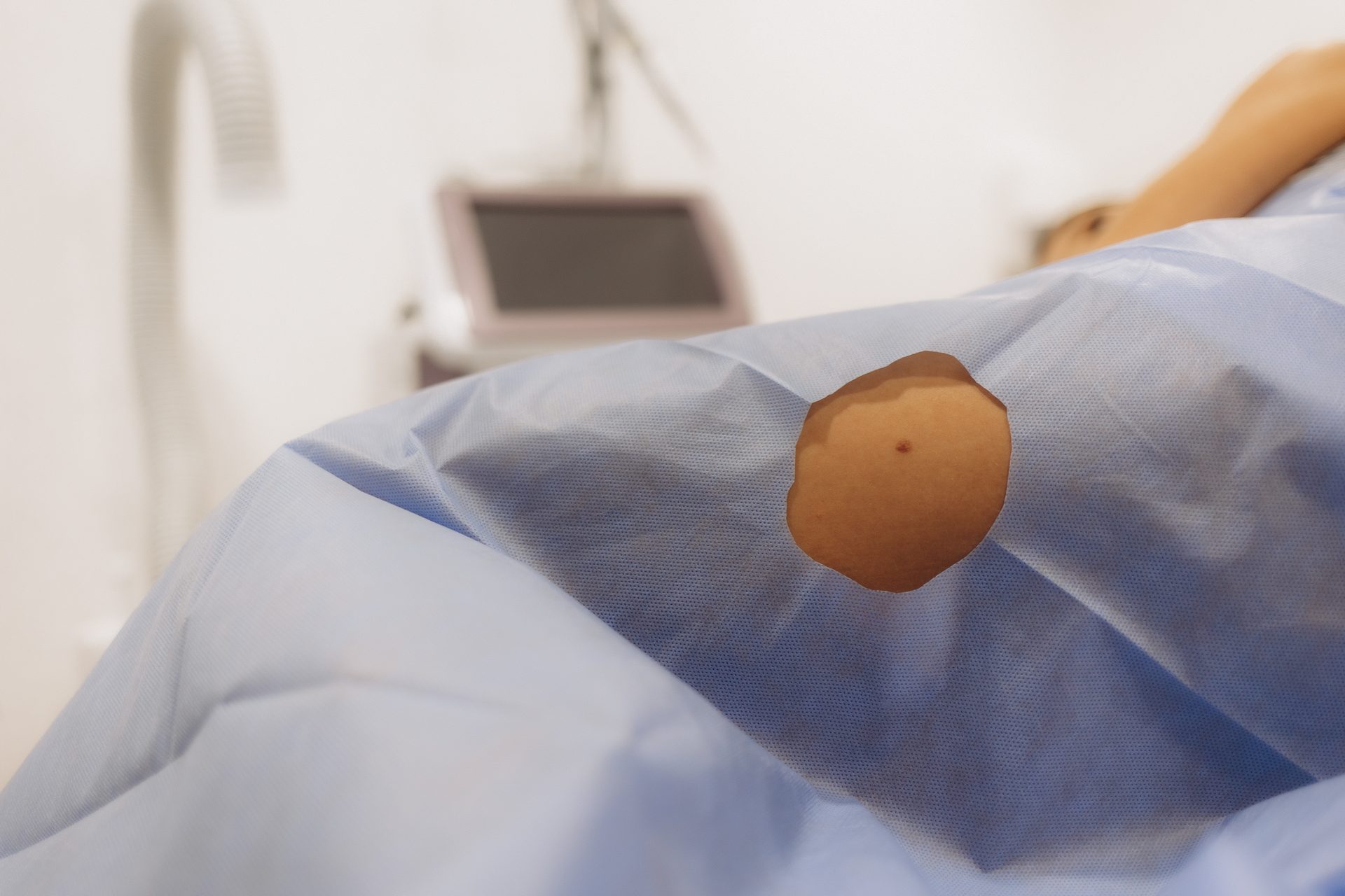
Skin cancer is the most common cancer in the United States. Most cases are highly treatable when found early. At Norton Dermatology & Wellness, a physician examines you in person, explains what they see, and decides with you whether a spot can be watched, photographed for comparison, or sampled. Nothing on this page replaces that conversation.
Names matter less than change. Still, it helps to know the language your physician may use.
Most common
Often a pearly, pink, or flesh-colored bump that can bleed, scab, or look like a sore that will not finish healing. It rarely spreads, but it can grow locally if left alone.
Sun-exposed skin
A scaly patch, firm bump, or tender horn-like growth — frequently on the face, ears, scalp, hands, or lips. It can grow more quickly than basal cell and should be examined promptly.
Least common, most serious
Can appear as a new mole or a change in an existing one. Early detection is the difference. If a spot is new, changing, or unlike your others, we want to see it.
Dermatologists use the ABCDE guide as a starting point — not a score you assign yourself. If any letter describes a spot, book a visit. One “ugly duckling” that looks different from the rest of your moles is also a reason to be seen.
Contact the practice or book the next available visit if you notice any of the following. If you are acutely unwell, use emergency care.
Anyone can develop skin cancer. We especially recommend a full-skin examination if you have:
Step 1
We ask what changed, when you first noticed it, and whether it itches, bleeds, or hurts.
Step 2
Scalp, face, trunk, limbs, nails, and other sites that are easy to miss at home.
Step 3
A lighted magnifier helps your physician see structure beneath the surface.
Step 4
Watch, photograph for comparison, treat in clinic, or biopsy — explained in plain language.
A biopsy is a small sample of skin, taken after the area is numbed. Your physician will tell you how the sample is taken, how to care for the site, and how results will be shared. Pathology interpretation belongs to your physician and the practice record. This website does not display biopsy results.
Current patients: if you are waiting on a result, or you were asked to return for treatment, sign in to the Patient Portal or call the desk. Do not wait on a changing spot because a previous visit was “probably fine.”
Once a skin cancer has been treated, we set a follow-up cadence that matches your risk — not a generic calendar. Self-checks at home still matter: once a month, in good light, including the scalp, back, and between the toes. Daily broad-spectrum sunscreen, shade, and avoiding tanning beds lower the chance of the next one.
For reserved follow-up time after a skin check, see Concierge in the header.
Reading about care is open to everyone. Booking a visit, reviewing a result, or sending a personal concern is private — those steps use the Patient Portal.
This page is educational. It is not a diagnosis, a treatment plan, or a substitute for a physician visit. If a spot is bleeding, rapidly changing, or you are worried today, contact the practice or seek urgent care.